
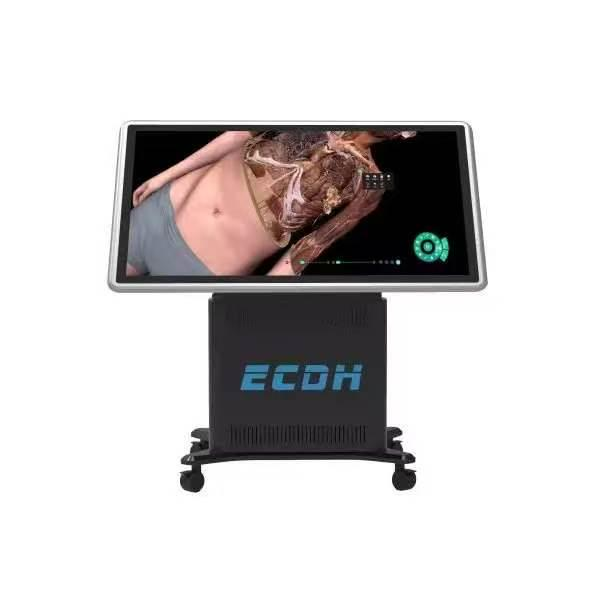
Medical training traditionally depends on physical specimens for anatomical instruction. While these traditional wet labs serve a fundamental purpose, they present continuous challenges for educational institutions. Universities face high recurring costs, difficult procurement processes, and complex storage requirements. Biological risks and toxic preservatives also limit student exposure outside scheduled hours.
Modern medical faculties now adopt advanced digital visualization platforms to resolve these logistical issues. By utilizing high-resolution, data-driven systems, educators can deliver clear, repeatable, and interactive training. These tools transform standard classrooms into dynamic learning environments without the physical constraints of traditional specimens.
Authentic Structural Data for Medical Simulation
Accurate instruction requires highly reliable source material. Foundational models used in modern digital platforms originate from real human tomographic imaging data rather than artistic approximations. For instance, systems developed by DIGIHUMAN utilize over 17,000 cross-sectional layers for male data and 16,000 for female data. The software renders each layer with resolutions reaching up to 13,700 by 6,340 pixels.
By harnessing tomographic imaging data with up to 1.2 billion original pixels, the platform achieves a 0.1 mm isotropic reconstruction accuracy. Every bone, nerve network, and muscular structure appears with strict adherence to human spatial relationships. By displaying over 6,000 specific anatomical structures in true three-dimensional form, institutions provide students with highly accurate anatomical models. This level of microscopic precision ensures that even subtle neural networks remain clearly visible to the user.
Enhancing Hands-On Interaction in the Laboratory
Students require active physical engagement to build spatial memory and understand structural relationships. A modern anatomy table facilitates this through intuitive touch controls. Users manipulate structures directly using simple, responsive gestures. One finger rotates the digital model, two fingers control the zoom function, and three fingers translate the image across the screen.
The software enables small student groups to perform virtual dissections, strip away fascia, isolate organ systems, and highlight specific tissue layers. Learners view the body from any angle, including internal perspectives looking outward from within thoracic or abdominal cavities. Hardware configurations featuring an 88-inch ultra-high-definition display with motorized height adjustment and 90-degree screen tilt support versatile classroom setups. Instructors easily transition between horizontal cadaveric orientation for group dissection and vertical display for lecture demonstrations. Students can also tag specific structures, record custom cutting planes, and save customized views to share with teammates during lab review sessions.
Transforming Pedagogy Through Active Case-Based Learning
Modern medical curricula rely heavily on Case-Based Learning (CBL) and Problem-Based Learning (PBL) to prepare students for clinical rotations. Rather than passively memorizing static atlas entries, student teams analyze real-world clinical scenarios directly within the classroom environment. Digital platforms support this approach by linking 3D structural models to extensive clinical libraries containing over 180 authentic patient cases, complete with chief complaints, diagnostic reports, and medical histories.
In a typical lab exercise, instructors assign different clinical presentations to student teams, such as acute appendicitis, vascular aneurysms, or complex bone fractures. Students compare pathological variations side by side with normal anatomical baselines, tracing structural deviations in real time. Integrated volumetric processing tools enable efficient loading and visualization of patient datasets, supporting smooth transitions between anatomy instruction and clinical case analysis. This interactive workflow encourages peer discussion, critical thinking, and early diagnostic reasoning, making abstract clinical concepts tangible for early-stage learners.
Bridging Preclinical Science and Multidisciplinary Training
Comprehensive medical education requires connecting macro-anatomy with microscopic histology and radiological imaging. Digital visualization platforms integrate cross-sectional CT and MRI data directly with 3D physical models. When examining a skull base or cardiac structure, students instantly toggle to matching radiological slices, gaining immediate proficiency in cross-sectional interpretation.
Classroom instruction also extends to developmental biology and micro-anatomy. Integrated embryology modules illustrate early human organogenesis through step-by-step animations, interactive quizzes, and structural timelines. For microscopic analysis, a digital slice library featuring over 2,000 histological sections allows students to zoom smoothly from 4X to 40X magnification. Instructors can seamlessly lead a class from gross organ dissection down to cellular structures within a single instructional session, allowing instructors to integrate gross anatomy and digital histology within a single teaching platform.
Optimizing Institutional Workflows and Faculty Efficiency
Replacing traditional wet labs with digital solutions dramatically streamlines institutional workflows and faculty management. Traditional dissection labs require specialized ventilation systems, continuous climate controls, and strict hazardous waste disposal protocols. Transitioning to digital dissection platforms eliminates recurring chemical expenses and minimizes facility maintenance demands while offering a safe, chemical-free learning environment.
To support multi-departmental usage across large institutions, advanced systems offer tailored user accounts and custom preset configurations. Faculty members create saved lesson plans, annotate specific 3D structures, and bookmark diagnostic histories for quick retrieval during lectures or practical examinations. High-performance computing configurations ensure fluid multi-user rendering across back-to-back classes. Whether serving undergraduate biology labs, nursing skill workshops, or postgraduate surgical anatomy courses, a single digital platform maximizes facility utility and long-term educational return.
Moreover, standardized digital assessment tools enable faculty to design practical quizzes with automated scoring, significantly reducing grading burdens while tracking student performance metrics over time.
